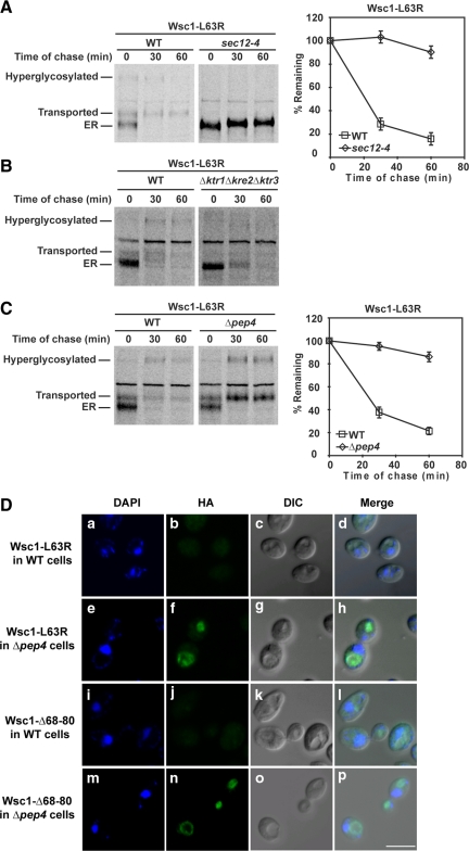
Figure 3.
Wsc1-L63R is transported to the vacuole via the Golgi for degradation. (A) Stability of Wsc1-L63R was examined in wild-type and sec12-4 cells using pulse-chase analysis as described in Figure 2A, except both strains were grown to log phase at 23°C and shifted to 37°C for 20 min before labeling. (B and C) Pulse-chase analysis was performed in wild-type, Δktr1Δkre2Δktr3, and Δpep4 cells expressing Wsc1-L63R as described in Figure 2A. (D) Wild-type and Δpep4 cells expressing Wsc1-L63R or Wsc1-Δ68-80 were fixed and permeabilized. Staining was performed using anti-HA primary antibodies followed by Alexa Fluor 488 goat anti-mouse secondary antibodies. DAPI staining marks the position of nuclei. Cells were visualized by confocal and DIC microscopy. Scale bar, 5 μm.

