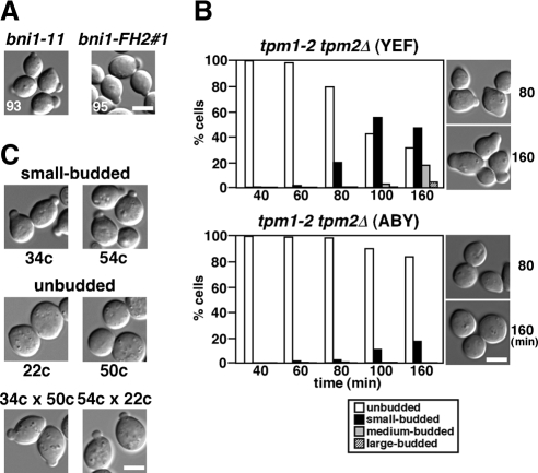
Figure 2.
The small-budded phenotype is common to other bni1-ts alleles and the tropomyosin mutant. (A) Growth arrest with a small bud in other bni1-ts bnr1Δ mutants. bni1Δ bnr1Δ strains (YKT1312 and YKT1313) harboring pRS314-bni1-11 and pRS314-bni1-FH2#1, respectively, were grown to early logarithmic phase and shifted to 37°C, followed by a 3-h incubation. Numbers indicate the percentage of small-budded cells. (B) Morphology of tropomyosin mutants with different genetic backgrounds. The α-factor–arrested tpm1-2 tpm2Δ (YEF) cells in the YEF473 strain background (YKT476) and the parental ABY944 tpm1-2 tpm2Δ (ABY; YKT286) cells were released into fresh medium at 37°C and fixed with 3.7% formaldehyde at the indicated time point. The graph shows the percentage of cells with the bud size that was categorized as described in Materials and Methods. Right, images of cells after incubation for the indicated time periods. (C) Morphology of bni1-116 bnr1Δ mutants with different genetic backgrounds. The bni1-116-EGFP bnr1Δ mutant (YKT533) in the YEF473 strain background was crossed with the tpm1-2 tpm2Δ (ABY) mutant (YKT286), and the resulting bni1-116-EGFP bnr1Δ progeny were morphologically examined. This allele of bni1-116 contains the C-terminally-fused EGFP with a drug resistance marker for convenience in tetrad analysis; we confirmed that the bni1-116-EGFP bnr1Δ mutant was indistinguishable from the bni1-116 bnr1Δ mutant in morphological and growth phenotypes at 37°C (data not shown). Exponentially growing cells were shifted to 37°C, followed by a 3-h incubation. Top and middle, images of representative progeny with small-budded (clones 34c and 54c) and unbudded (clones 22c and 50c) phenotypes, respectively. These morphologically different clones were crossed, and the resulting diploids were cultured as described above (bottom panel, 34c × 50c and 54c × 22c). Bars, 5 μm.

