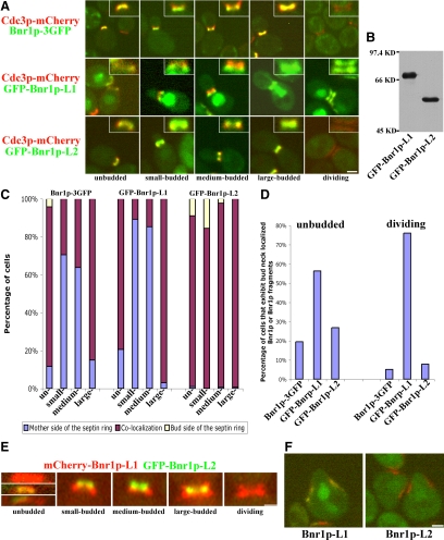
Figure 4.
Overexpressed GFP-Bnr1p-L1 and GFP-Bnr1p-L2 localize differently at the bud neck and in shmooing cells. (A) Localization of Bnr1p-GFP expressed from the BNR1 promoter and GFP-Bnr1p-L1 or GFP-Bnr1p-L2 expressed from the GAL1 promoter (after 2–3-h galactose induction) in cells expressing Cdc3p-mCherry through the cell cycle. On the top-right corner of each micrograph shows the enlargement of the neck region. (B) Equal amount of total lysates of strains overexpressing GFP-Bnr1p-L1 or GFP-Bnr1p-L2 were blotted with antibodies to GFP. (C) Quantitation of the localization of endogenous Bnr1p-GFP or overexpressed GFP-Bnr1p-L1 or GFP-Bnr1p-L2 with respect to septins through the cell cycle. (D) In unbudded or dividing cells where septins are present, percentage of cells that show colocalized Bnr1p-GFP or overexpressed Bnr1p-L1 or Bnr1p-L2. The data for C and D were obtained from one representative experiment with n ≥ 700 for each strain. (E) Localization of mCherry-Bnr1p-L1 and GFP-Bnr1p-L2 expressed from the GAL1 promoter, after a 2–3-h galactose induction. (F) Localization of GFP-Bnr1p-L1 or GFP-Bnr1p-L2 expressed from the GAL1 promoter in shmooing cells expressing Cdc3p-mCherry. Cells were induced with galactose for 2–3 h and treated with α-factor for 2 h.

