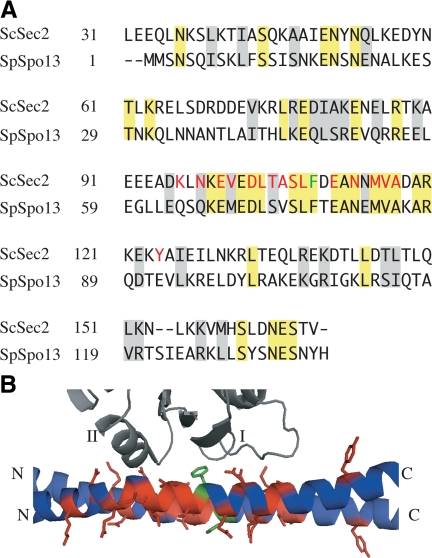
Figure 1.
SpSpo13 is homologous to the ScSec2 GEF domain. (A) Sequence alignment of ScSec2 GEF domain with SpSpo13. Identical residues are highlighted in yellow, and conservative changes are shaded gray. ScSec2 residues that are found in a close contact with ScSec4 in the cocrystal structure are in red (Dong et al., 2007). Spspo13 residues that are conserved in the ScSec4 binding surface are in green. Phe79 of SpSpo13 that was mutated to alanine is indicated in green. (B) View of the cocrystal structure of ScSec2 GEF domain and ScSec4 (PDB 2OCY; Dong et al., 2007). ScSec4 is in gray, above and the ScSec2 dimer is below. The switch I and II regions of ScSec4 are labeled. The side chains of the ScSec2 residues highlighted in A are shown in red, except for Phe109 in green.

