Figure 3.
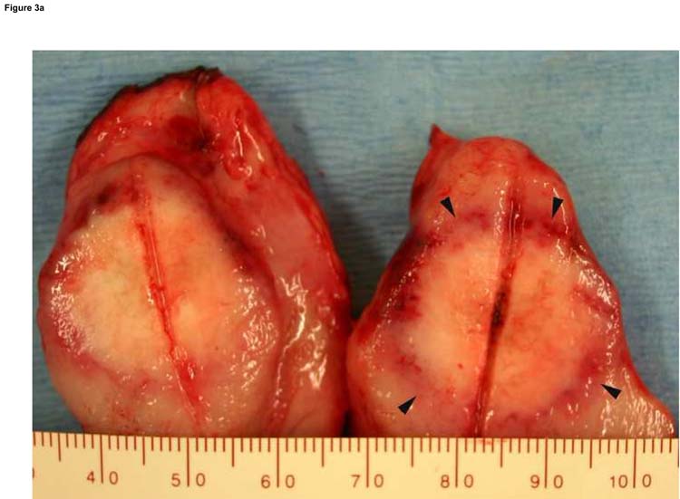
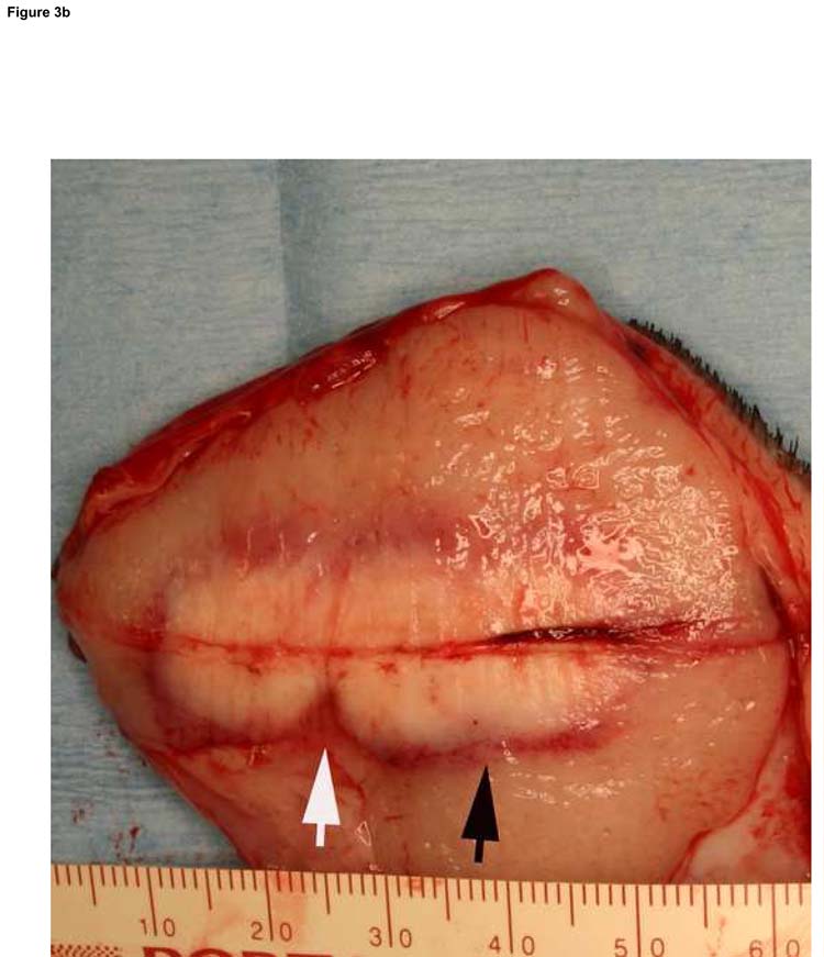
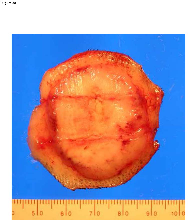
Typical ablation created with a single-applicator (a), single-exposure of 15 W for 120 seconds. The lesion shown (arrowheads) measures 20 mm × 23 mm in gross dimensions and contained an estimated thermally coagulated volume of 4987 mm3. Overlapping ablations were created by pull-back technique (b). Pulling back 1 or 1.5 cm resulted in contiguous ablation (black arrow) while pulling back 2 cm resulted in non-contiguous ablation zone (white arrow). Larger ablations were created with two applicators placed in parallel, separated by 1.5 cm (c). The ablation was performed with both fibers activated at 15 W for 120 seconds at two stations (total ablation time 240 seconds). The ablation zone measured 37 mm × 32 mm. Applicator tracks are seen as two parallel horizontal lines.


