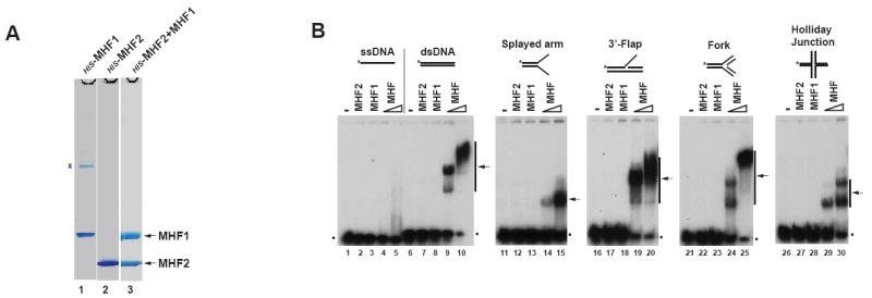
Figure 2. MHF binds double-strand and branch-structured DNAs, but not single-strand DNA.

(A) Coomassie blue-stained SDS-PAGE gels showing HIS-tagged recombinant proteins MHF1, MHF2 and MHF complex purified from E.coli. A nonspecific band was marked with “x”. (B) Electrophoretic mobility shift assay (EMSA) for testing DNA binding activity of MHF1, MHF2 and MHF complex. A variety of DNA substrates (0.5nM) illustrated at the top were incubated with 0.6 μM MHF1, 0.6 μM MHF2 and increasing amounts (0.3 and 0.6μM) of MHF complex, respectively. Asterisks denote 32P label at the DNA 5’ end. The arrows indicate the shift bands of MHF-DNA complex. The dots represent free DNA probe. (see also Figure S3).
