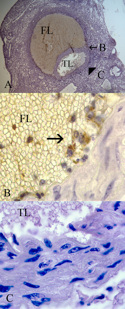
Figure 3.
Representative immunohistochemical analysis of an apolipoprotein E null mouse aorta harvested at 7 days of Ang II infusion. (A) At 7 days there is evidence of an aortic dissection with a false lumen (FL) and patent true lumen (TL) (20x magnification). Immunohistochemical staining for tyrosine-phosphorylated STAT1 was performed, and regions of the aorta bordering the false lumen (B) and the true lumen (C) were evaluated under higher magnification (100x). (B) Inflammatory cells infiltrating the false lumen stain positively (arrow) for tyrosine-phosphorylated STAT1, but no positive staining is observed along the true lumen border (C).

