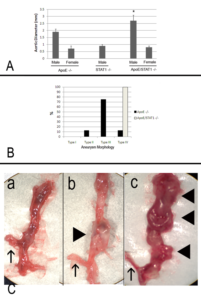
Figure 4.
(A) Aortic diameters from male and female ApoE −/−, STAT1 −/−, and ApoE/STAT1 −/− mice after 28 days of Ang II infusion. There were no significant aortic enlargements observed in STAT1 −/− male mice, nor in any of the female mice strains after 28 days of Ang II infusion. Aortic enlargement was observed in male ApoE −/− and male ApoE/STAT1 −/− mice, and were larger in the ApoE/STAT1 −/− mice compared to the ApoE −/− mice (P<0.05). (B) Graphic representation of aortic aneurysm morphology identified in ApoE −/− male mice (black bars) and ApoE/STAT1 −/− male mice. All ApoE/STAT1 null mice developed the type IV phenotype. (C) Examples of aortas resected from (a) an ApoE −/− mouse that did not undergo Ang II infusion, (b) an ApoE −/− mouse that developed a type II morphology aneurysm, and (c) an ApoE/STAT1 −/− mouse that developed a type IV morphology aneurysm. The upward pointing arrows in all three instances denote the location of the right renal artery, while the block triangles point to locations of aneurysmal degeneration (only b and c)

