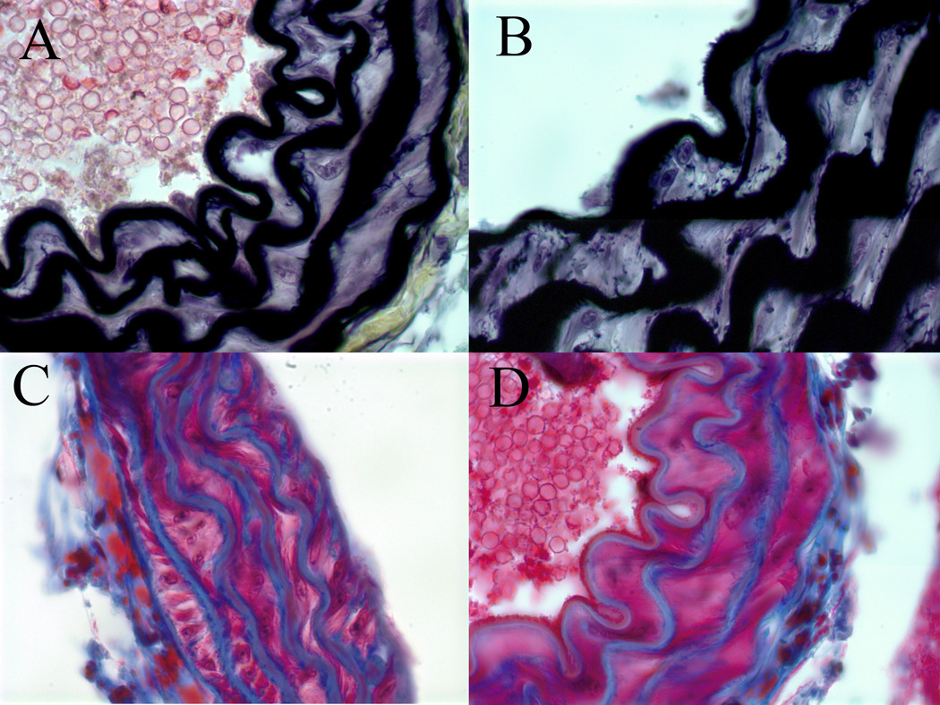
Figure 5.
Representative sections of histologic analysis of elastin (A, B) content with Movat stain, and collagen content (B, D) with trichrome stain from the aortic walls of apoE/STAT1 null mice (A,C) and apoE null mice (B,D) (100x). No differences in elastic lamellae, elastin deposition, or collagen deposition are seen at baseline between these two mouse strains.

