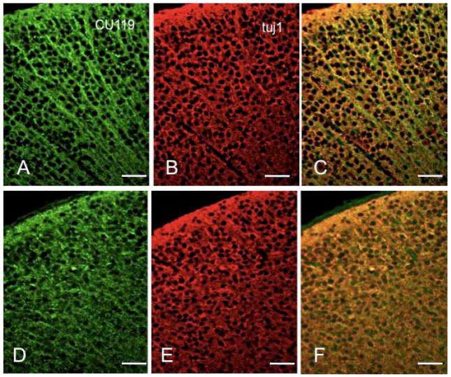
Figure 6.
MACF1 expression in the mouse cortex. Coronal sections of control (A–C) or Macf1 cKO (D–F) P0 brains were stained with CU119 polyclonal antibody (A, D), monoclonal tubulin antibody (B, E), and Hoechst (C, F). C, F, superimposed double images, with MACF1 staining in green and tubulin staining in red. Similar results were obtained from three pairs of animals from different litters. Scale bars: A–F=25μm

