
Figure 8.
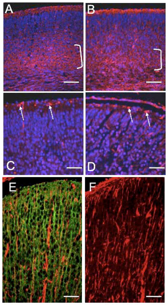
Cortical plate splitting and the radial glia are unaffected in Macf1 cKO brains. Coronal sections of control (A, C, E) or mutant (B, D, F) E17.5 brains were stained with CS-56 (A, B), reelin (C, D), or vimentin (E, F) antibodies (red), and co-stained with Hoechst nuclear dye (blue; A–D only) or CU119 antibody (green, E only). Brackets in A and B indicate subplate staining. Arrows in C and D indicate reelin-positive Cajal-Retzius cells in the marginal zone. E and F show vimentin-positive processes of radial glial cells, which did not colocalize with CU119-positive neuronal processes (E). Similar results were obtained for three pairs of animals from different litters. Scale bars: A–F=50μm