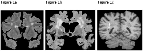
Figure 1. Illustration of MRI-markers of vascular brain injury.
a: MRI-scan (coronal T2-weighted sequence) of an 65-year old male participant with extensive white matter hyperintensities (EXT-WMH); b: MRI-scan (coronal T2-weighted sequence) of an 84-year old female participant with EXT-WMH; c: MRI-scan (coronal T1-weighted sequence) of an 84-year old male participant with an MRI-defined brain infarct in the right centrum semiovale (arrow)

