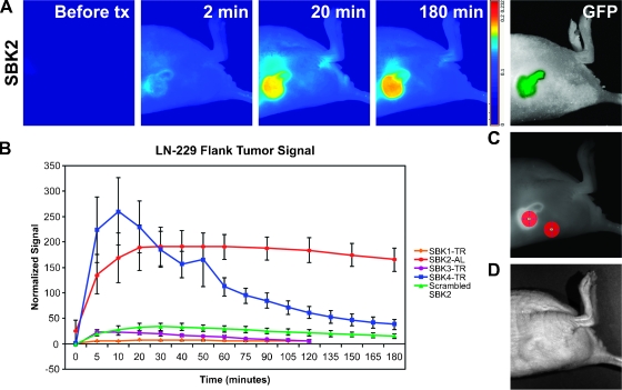
Figure 7.
PTPµ peptides SBK2 and SBK4 recognize LN-229 mouse flank tumors in vivo. TR- or Alexa-750 (AL)-conjugated PTPµ peptides were administered intravenously to mice with xenograft flank tumors of LN-229 cells. (A) Fluorescent images of SBK2-AL peptide labeling. Panel-labeled “before tx” (before time course) shows the animal autofluorescent background. The tumor cells are expressing GFP. (B) Time course of peptide binding to flank tumors (n = 3 animals tested per peptide). Average normalized signals acquired in the tumor region of interest were plotted. Error bars, SEM from the three animals. (C) ROIs shown over the tumor and nontumor skin. (D) Bright field image of the flank tumor labeled in A.

