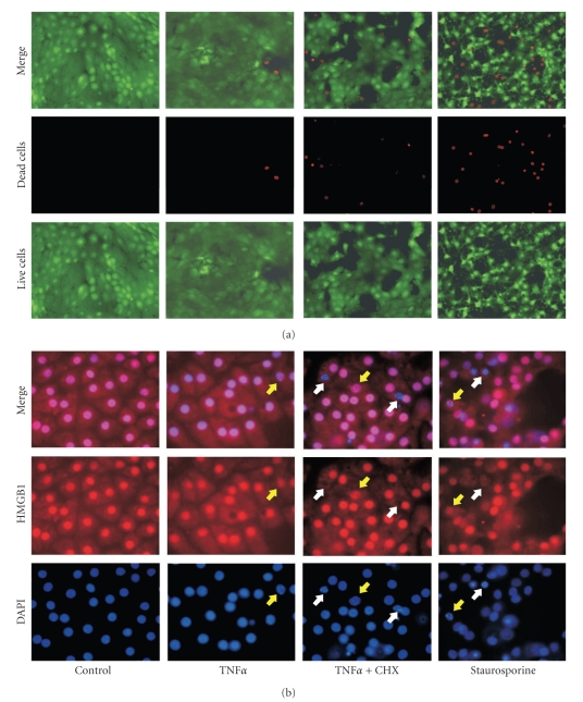
Figure 8.
TNFα induces HMGB1 release as well as HBGB1 binding in PCPEC. TNFα-stimulated PCPEC loose membrane integrity. PCPEC were stimulated with TNFα (100 ng/mL) ± cycloheximide (CHX) (1 μg/mL) for 10 hours. For a positive control PCPECs were treated with staurosporine (1 μM). (a) Adherent cells were labelled according to the Live/Dead assay protocol. Vital cells are penetrated by calcein and show a green fluorescence, damaged cells were penetrated by ethidium homodimer and show red fluorescent nuclei. Images were obtained by fluorescence microscopy (400 × magnification) using a digital camera. (b) Chromatin of vital and apoptotic cells binds HMGB1 (red) whereas TNFα ± CHX and staurosporine stimulated PCPEC partly released HMGB1 from nuclei (i.e., post-apoptotic necrosis; white arrows). DAPI staining revealed also that TNFα ± cycloheximide (CHX) resulted in chromatin condensation in PCPEC and binding of HMGB1 (i.e., apoptosis; yellow arrows). Both were detected by fluorescence microscopy (1000 × magnification) and photographs were taken using a digital camera. The whole well was examined for each experimental condition in two separate experiments; results from one representative experiment are shown.

