
Figure 3.
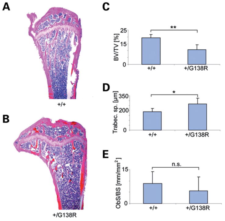
Skeletal phenotype of the ODDD-mutated mice. Comparison of wild-type (A) and Cx43+/G138R (B) tibial epiphysis and metaphysis shows more rarefied trabecular structure in the ODDD mutant relative to a wild-type littermate. Histomorphometric analysis reveals lower bone volume/total volume (**P < 0.001) (C), an increase in trabecular space (*P = 0.01) (D) and a non-significant (n.s.) decrease in osteoblast number (E) in mutants.