
Figure 5.
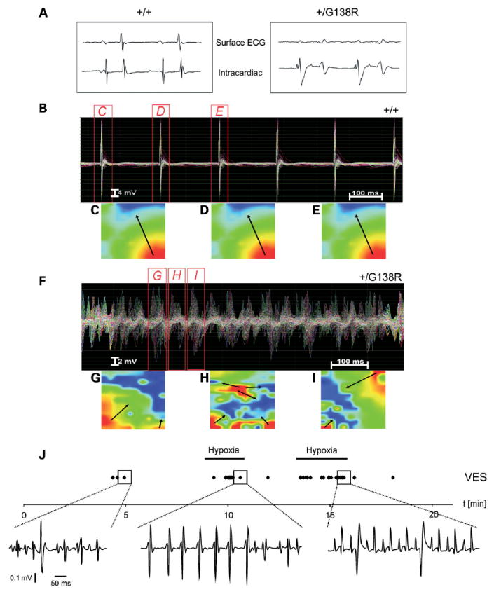
Disturbed impulse propagation and spontaneous cardiac arrhythmias developed in mutated hearts. (A) The disturbed impulse propagation in the ventricle is clearly seen in the striking broadening of the QRS complex in the surface ECG of the mutated hearts. (B) Normal impulse propagation and activation pattern in wild-type (n = 6) but ventricular tachycardias (VT) of all mutated (n = 8) hearts in Langendorff measurements. The cumulative Langendorff recordings of 128 epicardial electrograms clearly show repetitively directed and homologous conduction without signs of conduction disturbances or shift of conduction speed and direction in wild-type hearts during sinus rhythm (B–E) but a chaotic appearance of field potentials in the mutated ventricles during a polymorphic, high-frequency VT (F–I). Such polymorphic, incessant VTs were spontaneously present in all mutant hearts directly after the initiation of Langendorff extracorporal perfusion. EAMs of the left ventricular epicardium at three different time points during sinus beats in wild-type or polymorphic VTs in mutant hearts are shown in (C—E) or (G—I), respectively. The false color-coded reconstruction with red representing earliest and blue representing latest epicardial activation shows a heterogenic distribution of field potentials with inconsistent direction and speed of ventricular conduction in the Cx43G138R mutated mouse heart (arrows) during VT. This indicates different conduction pathways during different VT beats as typically present in polymorphic VT. (J) Cardiac arrhythmias recorded in vivo in cardiac-specific mutants. The frequency of the arrhythmias could be increased by hypoxia (hypoxia bars). Adjustment of O2 to normoxia largely abolished the arrhythmias. Arrhythmic events (VES) are displayed as individual points (VES during normoxia and under hypoxia). Even VT (1 VT ≥4 consecutive VES) could be recorded as a rare arrhythmic event during hypoxia (middle panel).