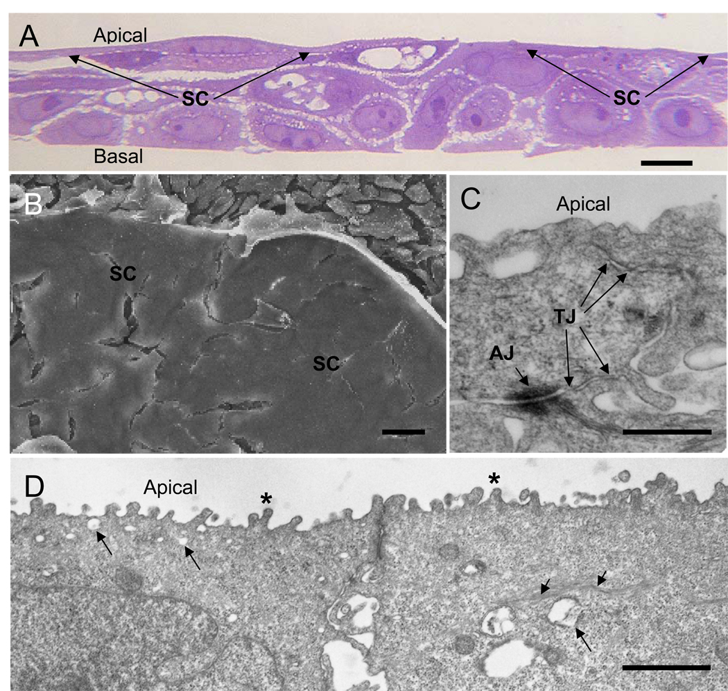
Figure 1.
TEU-2 cells differentiate into a stratified epithelial culture consisting of thin, tightly apposed apical superficial cells and more loosely connected underlying cells. (a) Light micrograph of cultured TEU-2 cells stained with toluidine blue. Apical and basal surfaces are indicated and the superficial cell (SC) layer is indicated with arrows. (b) SEM image of the apical surface of TEU-2 cell culture. The apical superficial cell (SC) surface appears flat and featureless. The loosely connected underlying cells are apparent where the SC layer has separated from the underlying cells during processing. (c) TEM of apical tight junctions (TJ) and an adhering junction (AJ) between two SCs. (d) Lower magnification TEM of SCs illustrating the stubby microvilli (asterisks), the clear cytoplasmic vesicles (long arrows), the strands of tonofilament fibers (short arrows) and adhering junctions. Magnification bars in a, b, c and d are 10, 10, 0.5 and 1 µm, respectively.

