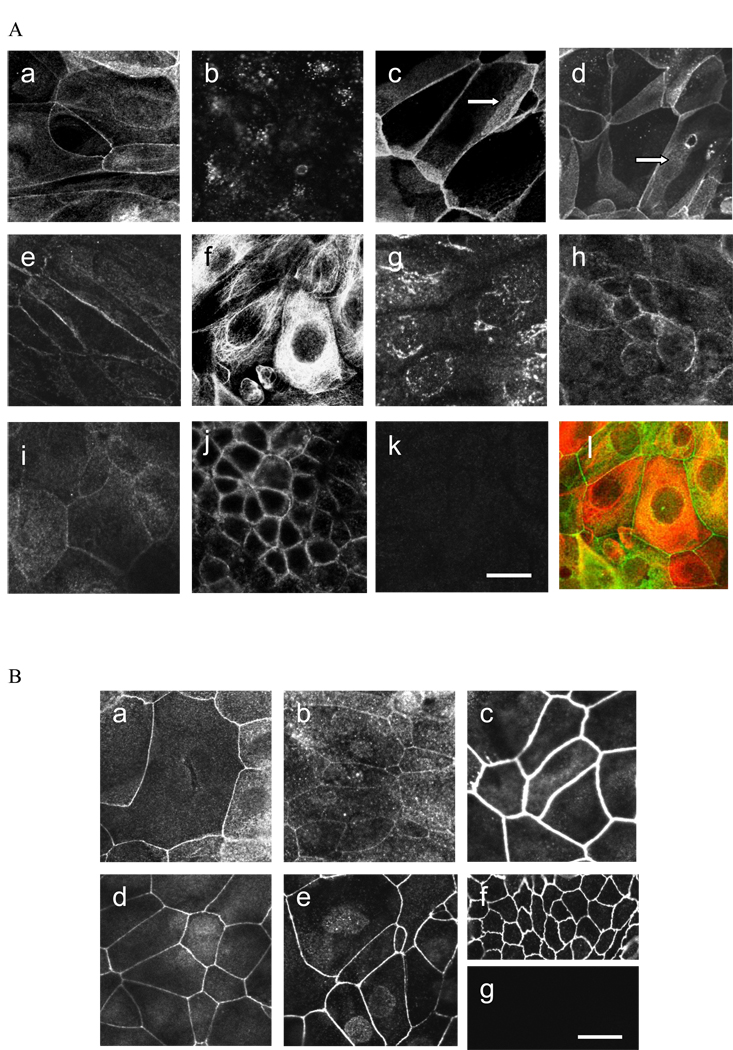
Figure 2.
(A) Expression of claudins in TEU-2 cells: (a) claudin 1, (b) claudin 2, (c) claudin 4, (d) claudin 5, (e) claudin 7, (f) claudin 8, (g) claudin 12, (h) claudin 14, (i) claudin 16. Arrows in (c) and (d) indicate claudin immuno-staining along apical-lateral cell borders between overlapping cells. (j) Positive control staining for claudin 4 in Caco-2 cells. (k) Representative example of negative control. Normal rabbit IgG for claudin 4. (l) Merged double-labeled image of claudin 8 (red) and ZO-2 (green). (B) Expression of TJ-associated proteins in TEU-2 cells: (a) occludin, (b) JAM-1, (c) ZO-1, (d) ZO-2, (e) ZO-3. (f) Representative positive control for ZO-3 in MDCK cells. (g) Representative negative control of TEU-2 superficial cells stained with normal rabbit IgG. Magnification bar = 25 µm.

