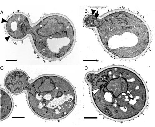
Figure 5.
The sec3Δ cells accumulate secretory vesicles and exhibit mislocalized endoplasmic reticulum. Wild-type (A and B) and sec3Δ cells (C and D) grown in SC medium at 25°C were processed for electron microscopy as described in the MATERIALS AND METHODS. Bar, 1 μm. Black arrowheads point to cortical ER in the daughter cells. Note the absence of cortical ER in the large bud of the sec3Δ cell (C). ER tubules extending into the bud (arrow) can be seen in B. An example of ER accumulation in the mother of a sec3Δ cell is shown in D (white arrowhead).

