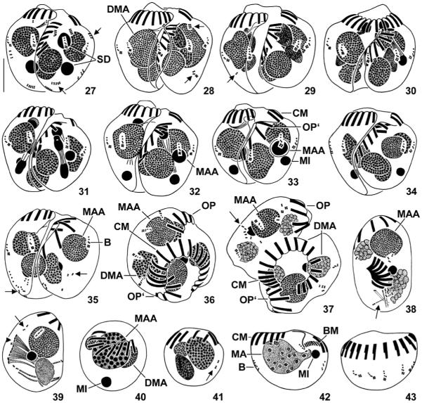
Figs 27–43.
Halteria grandinella, conjugants during (27–32) and after (33–37) second synkaryon division and exconjugants (38–43) after protargol impregnation. 27–29. Ventral view of pairs during interphase, prophase, and metaphase. Note the short and faintly impregnated rows of granules (arrows; probably somatic kinetids). 30–32. Dorsal (30) and ventral (31, 32) view of pairs during anaphase (31) and telophase (30, 32). The posterior division product of the more anterior derivative becomes the macronucleus anlage, the posterior division product of the more posterior derivative represents the developing micronucleus; their sister derivatives disintegrate. 33–35. Ventral view of pairs during late stages of conjugation. Occasionally, scattered somatic kinetids (arrows; 35) occur. 36, 37. Top view of pairs just before separation of conjugants. Note the slightly disordered somatic ciliature (arrow; 37). 38–43. Exconjugants. The globular specimens (40, 41) might not be viable. Arrows (38, 39, 41) mark scattered somatic kinetids. Arrowhead (39) denotes forming or disintegrating membranelles. B – bristle kineties, BM – buccal membranelles of exconjugant, CM – collar membranelles of right conjugant or exconjugant, DMA – degenerating fragments of vegetative macronucleus, MA – new vegetative macronucleus, MAA – macronucleus anlage, MI – developing or mature micronucleus, OP – oral primordium of right conjugant, OP’ – oral primordium of left conjugant, SD – synkaryon derivatives. Scale bar 10 μm.

