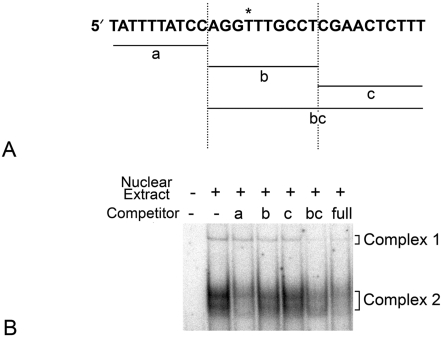
Figure 8. Complex 2 is formed by DNA–protein interactions involving nucleotides upstream of the polymorphic site.
(A) Shown is the 30 bp sequence delineating unlabelled competitor DNAs for the 129B6 polymorphic region. The polymorphic site is denoted by an asterisk. Fragments a, b and c span the individual regions shown. These fragments were used for competition as concatemers of three copies of each 10 bp region. (B) Representative PhosphorImager-generated autoradiograph of unlabelled co-competition experiments with different competitor DNAs reacting with 129B6 retinal extract. The radiolabelled 129B6 full-length probe was incubated in the presence or absence of unlabelled competitor DNAs. Each competitor DNA was used at a 150-fold molar excess of potential binding sites. Competitor a specifically competed Complex 2, while b competed Complex 1, but minimally competed Complex 2. Alternatively, oligo bc efficiently competed Complex 2. These results suggested Complex 2 was formed with interactions upstream of the polymorphic site, but may also involve interactions further downstream of this site.

