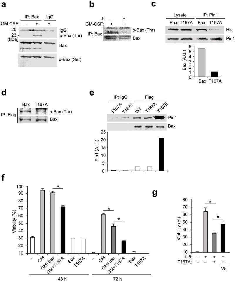
Figure 4. Phosphorylation of Bax Thr167 facilitates cell survival.
(a) Eosinophils were left untreated (–) or incubated for 4 h with GM-CSF. Cells were lysed in CHAPS buffer and immunoprecipitated (IP) with anti-Bax or nonimmune IgG, followed by immunoblot with anti-p-Thr, anti-p-Ser and anti-Bax. (b) Eosinophils were treated with GM-CSF alone or together with juglone as in (a). Cell lysates were immunoprecipitated with anti-Bax prior to immunoblot with anti-p-Thr and anti-Bax. (c) Eosinophils were incubated with His-tagged TAT-WT-Bax (Bax) or TAT-T167A-Bax (T167A) for 10 min in the presence of GM-CSF. Cell lysates were pre-cleared and immunoprecipitated with anti-Pin1 followed by immunoblot with anti-His and anti-Pin1. Top, representative immunoblot. Bottom, the density of Bax and T167A bands, normalized to the Pin1 bands. (d) Eosinophils were incubated with Flag-tagged TAT-WT-Bax or TAT-T167A-Bax. After treatment with GM-CSF, cell lysates were immunoprecipitated with anti-Flag followed by immunoblot with anti-p-Thr and anti-Bax. (e) WT, T167A and T167E Bax proteins were incubated in vitro with GST-Pin1 in CHAPS buffer prior to immunoprecipitation with anti-Flag or nonimmune IgG followed by immunoblot with anti-Pin1 or anti-Bax. Top, representative immunoblot. Bottom, the density of Pin1 bands, normalized to the Bax bands. (f) Eosinophils were incubated with or without Flag-tagged TAT-WT-Bax or TAT-T167A-Bax in the presence or absence of GM-CSF (50 pg/ml). Cell viability was determined after 48 and 72 h. (g) Cells were incubated with or without IL-5 (100 pM) alone or together with TAT-Bax-T167A or V5. Cell viability was determined after 72 h). *, P < 0.05 Student’s t-test in a two-tailed analysis. Immunoblots are representative of at least 3 experiments with different donors.

