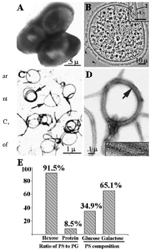
Fig. 1.

Electron micrograph of intact Pn6A bacteria (A) and cross-sectioned Pn6A bacterium (B) as well as purified peptidoglycan-polysaccharides (PGPS) after treatment with 8% sodium dodecyl sulfate at 100°C for 15 minutes (C, lipid dissolved); 1 = plasma membrane; 2 = cell wall; c = capsular layer. (D) Amplification of C, with insert indicating that peptidoglycan moiety of Pn6A contains filament-like structures (peptidoglycan backbone). Note that polysaccharide moiety of Pn6A is not visible under electron microscopy. Biochemical analysis indicates that ratio of carbohydrate-to-protein of PGPS is approximately 11:1 (91.5% carbohydrates and 8.5% proteins by hexose and protein assays, E). Carbohydrate compositions are mainly glucose (34.9%) and galactose (65.1%) with 1,2- and 1,4-glycosidic linkage as judged by gas chromatography/mass spectrometry.
