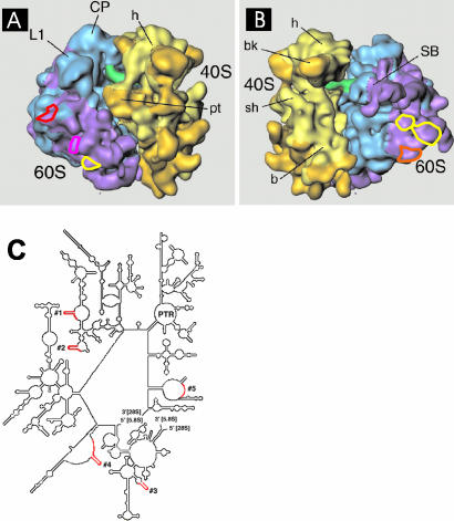
Figure 1.
Targeted hybridization sites on the 60S ribosomal subunit. (A and B) Colored circles show the locations of the expansion sequences for which complementary oligonucleotides probes were designed. (A) Yellow: oligo 1; pink: oligo 2; red: oligo 3. (B) Yellow: oligo 4; orange: oligo 5 (see MATERIALS AND METHODS for sequences of each oligo). The cryo-EM maps of the S. cerevisiae 80S ribosome are taken from Spahn et al. (2001) and reproduced with permission of CELL Press (Cambridge, MA). (C) 28S secondary structure map (yeast) showing regions targeted for oligo hybridization in red.

