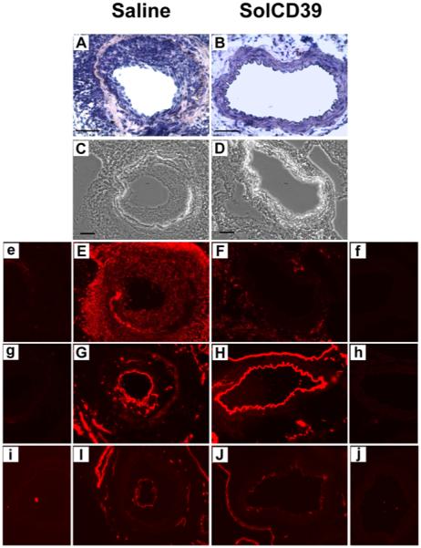
Fig. 4. Immunohistochemical analysis of mouse femoral arteries 19 days after wire-induced endothelial injury.

Immunohistochemical analysis was performed on representative cross-sections of femoral arteries of mice injected with human solCD39 or saline to identify specific cell types. Sections were stained with hematoxylin-eosin (A, B; 40x) as well as visualized under bright-field illumination (C, D; 20x). Sections were also immunofluorescently stained for leukocytes (E, F; anti-CD45 antibody, 20x), and EC (G, H; anti-CD31 (PECAM-1) and I, J; anti-panendothelial cell antigen antibodies, 20x). Images of the corresponding IgG isotype control to each specific antibody are labeled with lower case letters (e-j). Bar = 50 μm.
