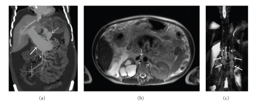
Figure 1.
(a) Coronal plane maximum-intensity-projection reformatted CT image shows the fusiform dilatation of the main portal vein (thick arrow). There is also marked dilatation and tortuosity at coronary vein (thin arrow). Note that the caliber of superior mesenteric vein is normal (arrowhead). (b) Axial half-Fourier acquisition single-shot turbo spin-echo (HASTE) MR image demonstrates thick lymphatic channels in the retrocrural space (arrows). Also, there is hydnonephrosis at the right kidney. (c) MR-Lymphangiography image (a half-Fourier single-shot turbo spin-echo 2D sequence with breath-hold technique with maximum intensity projection (MIP)) demonstrates two tortuous tubular structures on each side of the aorta representing dilated cisterna chyli (thin arrows). The meshwork of saccular lymphatic channels in the lumbar region inferior to the cisterna represents the dilated lumbar lymphatics (thick arrows).

