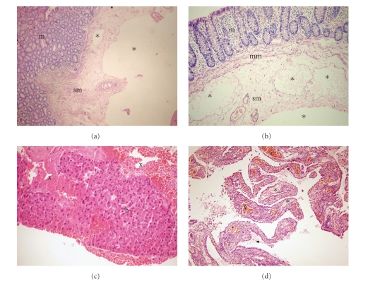
Figure 2.
(a), (b) Edema of the submucosa and muscularis mucosa, and dilatation of the submucosal lymphatic vessels (asterixes). (c) Blood filled cavities (x) in the liver biopsy, (d) peritoneal biopsy displaying dilated lymphatic (asterixes) and capillary (v) vessels. m: mucosa, sm: submucosa, mm: muscularis mucosa, h: hepatocytes.

