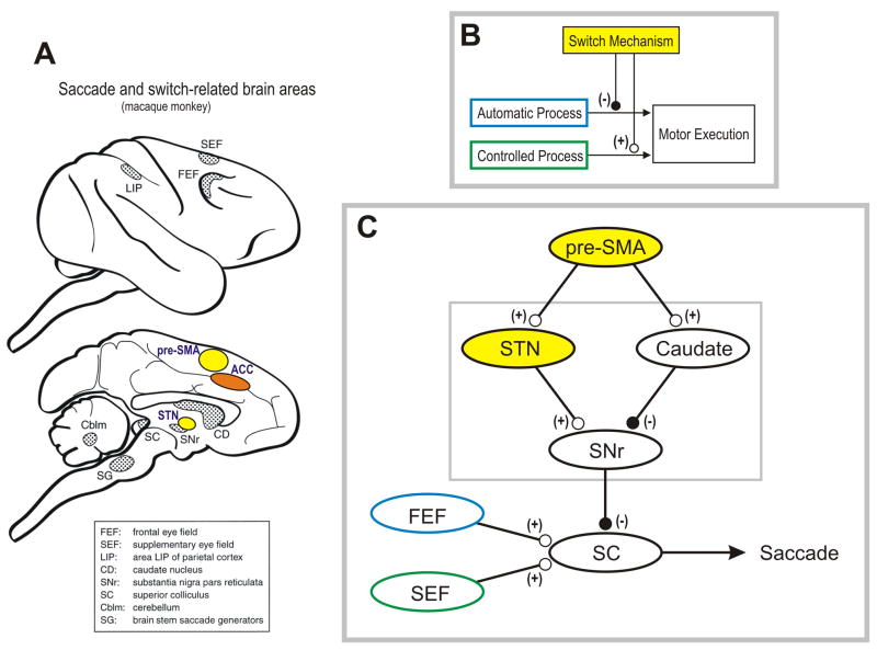
Figure 2. Neural mechanism of proactive switching in oculomotor behavior.
A neural mechanism of behavioral switching must be able to (1) detect a change in the context, (2) suppress the prepotent, automatic process, and (3) facilitate the alternative, controlled process (conceptual scheme). The suppression must occur quickly because the automatic process emits a motor signal quickly; the facilitation can occur thereafter because the controlled process is slow. Recent studies have suggested that the pre-SMA, together with other frontal cortical areas, acts as a switch mechanism and the basal ganglia may mediate the switch-related signal from the cortical areas. In our study using saccadic eye movement, many neurons in the pre-SMA became active selectively and proactively on switch trials (Box 2). It was also shown, using a go-nogo task, that some pre-SMA neurons suppress the prepotent saccade, others facilitate the alternative saccade, and the rest have both functions. The suppressive pre-SMA neurons tended to be active earlier than the facilitatory pre-SMA neurons, consistent with the conceptual scheme. In the basal ganglia, the STN may serve to suppress the automatic saccade by enhancing the inhibitory output of the basal ganglia (SNr) on the SC or the thalamo-cortical network. The caudate nucleus might serve to facilitate the controlled saccade by disinhibiting the target of the basal ganglia. We speculate that the signals for the automatic and controlled saccades are carried mainly by the frontal eye field (FEF) and the supplementary eye field (SEF) respectively. In the possible neural network, excitatory and inhibitory connections are indicated by (+) and (−) respectively.

