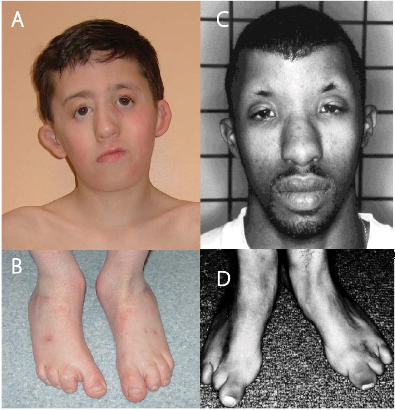
Figure 1. Clinical characteristics of an individual with Miller syndrome (A,B) and an individual with methotrexate embryopathy (C,D).

A 9 year-old boy with Miller syndrome (A and B) caused by mutations in DHODH. Facial anomalies (A) include cupped ears, coloboma of the lower eyelids, prominent nose, micrognathia and absence of the 5th digits of the feet (B). A 26 year-old man with methotrexate embryopathy (C and D). Note the cupped ears, hypertelorism, sparse eyebrows, and prominent nose (C) accompanied by absence of the 4th and 5th digits of the feet (D). C and D are reprinted with permission from Bawle et al. Teratology 57:51-55 (1978).
