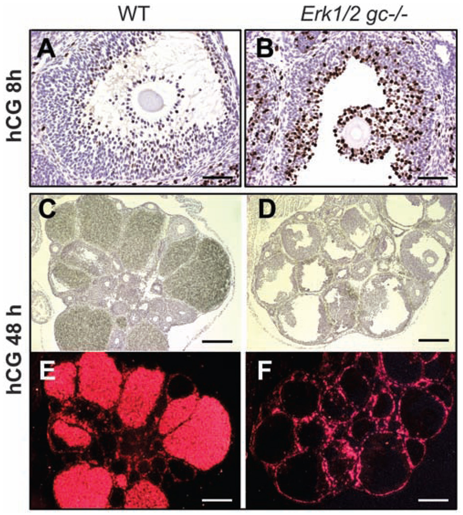
Fig. 2.
ERK1/2 are required for the terminal differentiation of GCs during ovulation. (A and B) BrdU staining of WT and Erk1/2gc−/− ovary sections at 8 hours after hCG treatment. Scale bars, 62.5 µm. (C to F) In situ hybridization shows the expression of Cyp11a1 mRNA in ovaries of WT and Erk1/2gc−/− mice at 48 hours after hCG treatment. Histology of the ovaries is shown by hematoxylin staining (C and D); localization of Cyp11a1 mRNA is shown by dark-field images (E and F). Scale bars, 250 µm.

