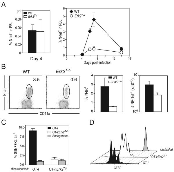
FIGURE 6.
Erk2 is required for CD8 T cell survival following infection with VSV. A, WT or Erk2T−/− CD8 T cells were infected i.v. with VSV, and Ag-specific CD8 T cell responses in peripheral blood were assayed at indicated time points using H-2Kb-RGYVYQGL tetramers. Percentages of PBL that are tetramer-positive are shown. The data presented are the means ± SD and are representative of two independent experiments. B, Representative FACS plots of WT and Erk2T−/− mice (gated on CD8+ cells), percentage, and total numbers of Ag-specific cells present in spleen on day 6 postinfection. The numbers shown in the FACS plots are percentage of total lymphocytes, and the bar graphs depict the mean ± SD for a cohort of mice. The data presented are representative of two independent experiments. C and D, Congenically marked (CD45.1+) naive mice received an i.v. transfer of 1000 unlabeled (C) or 0.5 × 10⁶ CFSE-labeled (D) WT or Erk2T−/− OT-I cells 1 day before immunization with VSV-OVA. Spleens were isolated at day 2 (D) or day 6 (C) postinfection and analyzed by FACS for the presence of OVA-specific donor (OT-I) and host (endogenous) CD8 T cells. The graphs presented in C are expressed as a percentage of total lymphocytes in the spleen, and the plots in D are gated on donor OT-I cells (CD45.2+CD8+). Also indicated in D are the levels of CFSE within undivided cells. The data are representative of two independent experiments.

