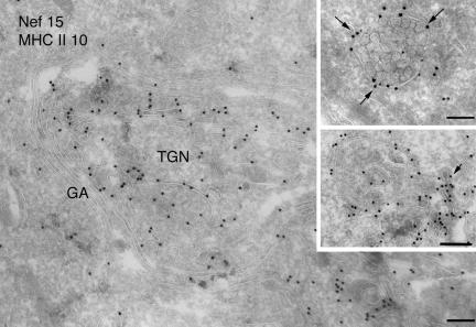
Figure 5.
Immunoelectron microscopy analysis of Nef-expressing cells. Ultrathin cryosections of HeLa-CIITA cells transfected with Nef-FT were double immunogold-labeled with the anti-Nef mAb MATG (protein A gold [PAG] 15) and the anti-DR polyclonal antibody (PAG 10). Bar, 200 nm. Nef is associated with numerous membrane vesicles and tubulovesicular elements at the TGN. The Nef protein is also associated with the cytosolic side of the limiting membrane of MVB (top inset, arrows) and closely apposed tubulovesicular structures (bottom inset) that are often coated (see arrow). Bar (insets), 100 nm.

