Figure 3.
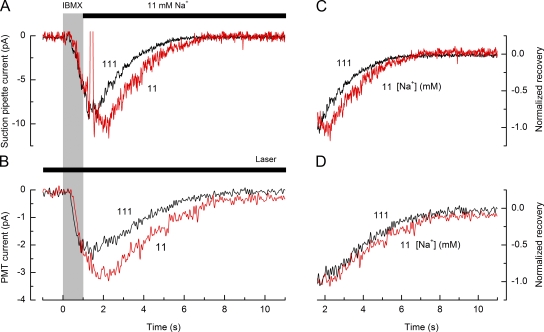
Effect of external Na+ concentration on receptor current recovery. (A) Suction pipette recordings of receptor current from an isolated ORN in response to a 1-s stimulation with 200 µM IBMX. Afterward, the cilia were rapidly returned to normal Ringer’s solution with 111 mM Na+ (black trace) or to a partially guanidinium-substituted Ringer’s solution containing 11 mM Na+ (red trace). Gray bar denotes the command for the presentation of the IBMX stimulus. Solid bar denotes exposure to partially guanidinium-substituted Ringer’s solution with an 11-mM Na+ concentration. (B) PMT current evoked by the change in ciliary Ca2+ dye fluorescence recorded simultaneously with the suction pipette current traces shown in A. The Na+ concentration in the Ringer’s solution is indicated beside each trace. Suction pipette current traces have been junction corrected, and each trace represents the average of two to three trials. PMT current traces have been low-pass filtered at 10 Hz and baseline corrected to the fluorescence level before stimulus onset. Solid bar denotes laser excitation of fluorescence. (C and D) Recovery phases of the suction pipette and PMT current traces from A and B after normalization to their peak magnitude for the comparison of their time course.

