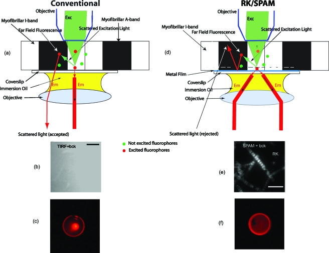
Figure 1.
Concept of SPAM microscope. A cardiac myofibril is illuminated from above. (a) In a conventional microscope all light, including scattered (background) light, is able to penetrate the coverslip. Green dots represent fluorophores that are out of the field of excitation. Red dots represent fluorophores that are in the path of direct or scattered excitation light. (d) In RK/SPAM, a sample is placed on a metal-coated coverslip and excited with green light (right). The excitation energy from the excited fluorphore couples to the surface plasmons and radiates through the metal film (red) to the objective as a surface of a cone with a half angle equal to the SPCE angle. Metal can be a thin layer of Al (20 nm thick), or Ag or Au (50 nm thick). The scattered light is unable to penetrate the coverslip and is radiated into free space. (b) and (e) The background rejection by SPAM. 0.5-mM rhodamine 800 added as background obscures the image in ordinary TIRF (b). SPAM in the RK configuration eliminates much of the background contribution (e). Myofibrils (0.1 mg∕mL) were labeled with 100-nM Alexa647-+10-μM unlabeled phalloidin for 5 min at room temperature, then extensively washed with rigor buffer containing 50-mM KCl, 2-mM MgCl2, 1-mM DTT, 10-mM TRIS pH 7.0. 633-nm excitation, 1.65 NA 100× Olympus objective, sapphire substrate, 1.78 refractive index immersion oil. The bars are 5 μm in (b) and 10 μm in (e). (c) and (f) The back focal plane image of a sample in a microscope in (c) conventional configuration consists of weak outer ring and a diffuse interior with s strong center corresponding to imperfect blockage of exciting light. The BFP image of a sample in a microscope in (f) RK configuration consists of a strong ring corresponding to emission into free space in a cone.

