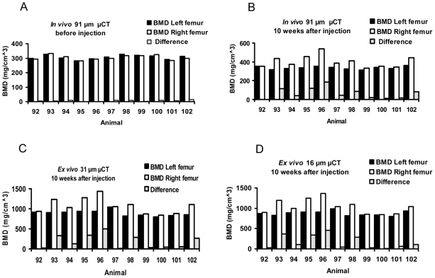
Figure 2. Bone mineral density (BMD) of the diaphysis was greater in the femora involved by prostate cancer.
Diaphyseal BMD was measured in both femora by in vivo μCT before injection (A) and 10 weeks after injection (B) of vehicle (left femora) or prostate cancer cells (right femora), and then by ex vivo specimen μCT at 31 µm (C) and 16 µm (D) voxel sizes.

