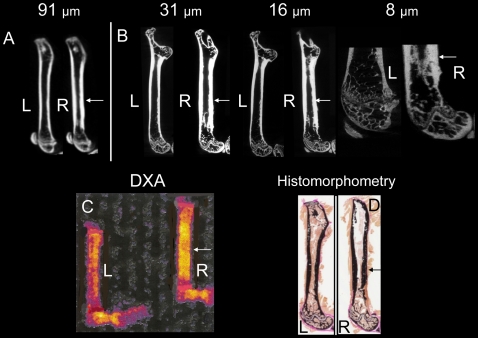
Figure 3. Representative in vivo and ex vivo images demonstrate morphologic changes of mineralized cortical and trabecular bone induced by prostate cancer involving the skeleton.
Sagittal in vivo μCT and ex vivo specimen μCT images of femora 10 weeks after injection of vehicle (left) or prostate cancer cells (right). (A) In vivo μCT (91-µm voxel size); (B) ex vivo specimen μCT at 31-µm, 16-µm, and 8-µm voxel sizes; (C) ex vivo dual-energy X-ray absorptiometry (DXA); and (D) bone histomorphometry images are presented. L, left femur; R, right femur with tumor; arrow, cortical thickening.

