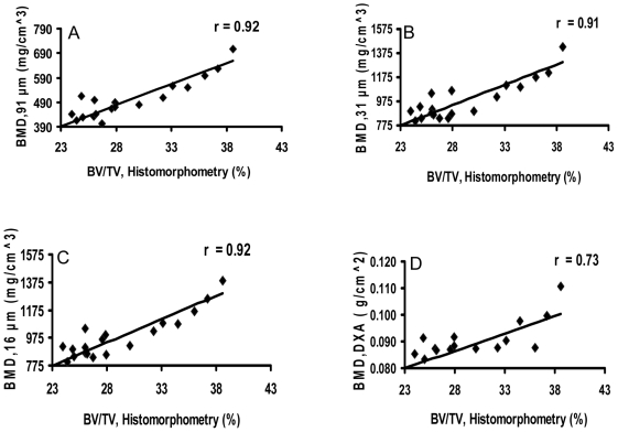
Figure 5. In vivo μCT, ex vivo specimen μCT, and DXA measurements of diaphyseal bone mineral density (BMD) correlate with histomorphometry.
In vivo μCT (91-µm, A), ex vivo specimen μCT (31-µm, B; 16-µm, C), and DXA (D) measurements of bone mineral density (BMD) correlate with histomorphometric measurements of mineralized bone volume/total bone volume (BV/TV).

