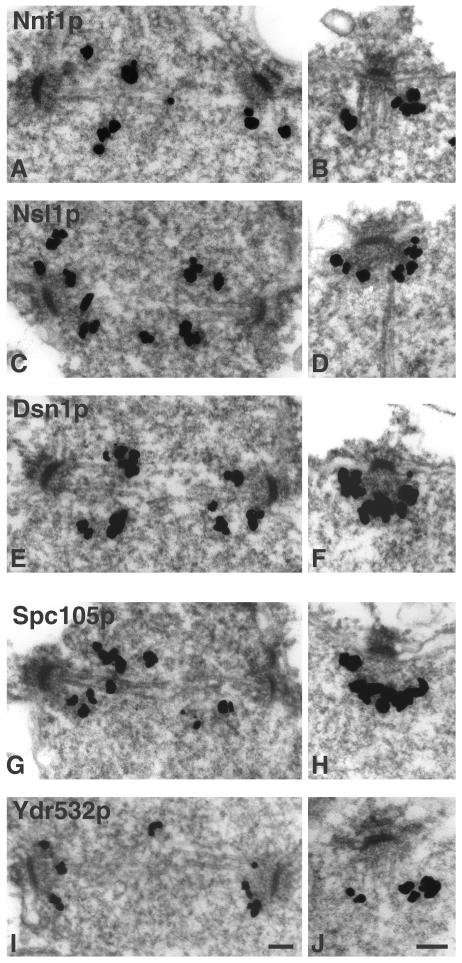
Figure 3.
Immuno-EM of GFP-tagged proteins in the indicated strains. (A and B) Nnf1p (VNY3), (C and D) Nsl1p (VNY50), (E and F) Dsn1p (VNY28), (G and H) Spc105p (MSY52), and (I and J) Ydr532p (JK1495). Staining is seen between SPBs in short spindles (left side) and in association with the nuclear face in all other SPBs (right side). Bar, 0.1 μm.

