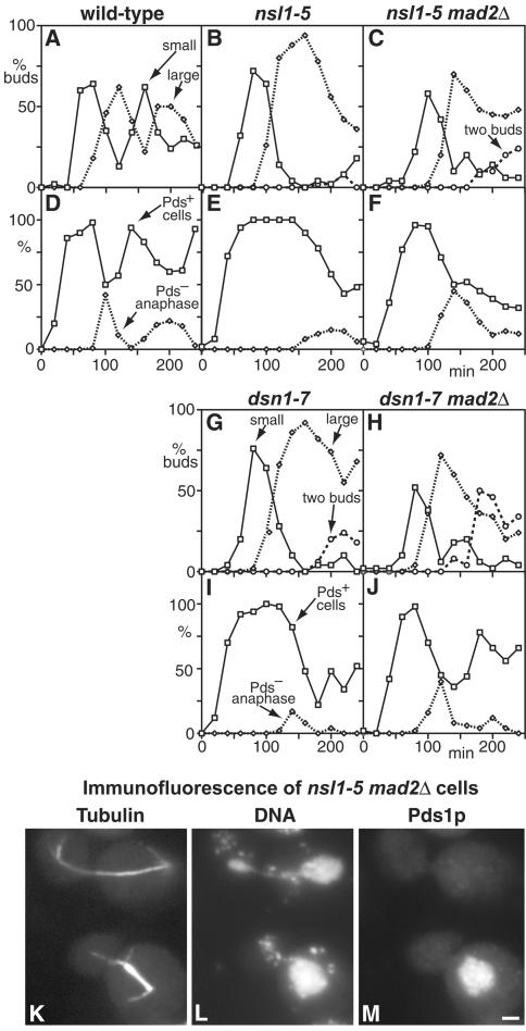
Figure 7.
The arrest of nsl1-5 and dsn1-7 mutants during mitosis is Mad2p-dependent. (A–J) Wild-type (A and D; K6445), nsl1-5 (B and E; VNY130), nsl1-5 mad2Δ (C and F; VNY318), dsn1-7 (G and I; VNY116), and dsn1-7 mad2Δ (H and J; VNY283) cells were synchronized in G1 with α-factor and released at 36°C. The percentage of cells with small buds, large buds, or two buds was plotted against time (A–C, G, and H), as were the percentages of Pds1p-positive cells and Pds1p-negative cells containing anaphase spindles as determined by immunofluorescence (D–F, I and J). (K–M) Immunofluorescence of nsl1-5 mad2Δ cells stained with antitubulin (K), DAPI (L), and antimyc for Pds1p (M). Pds1p-negative cells with anaphase spindles show unequal DNA segregation. Bar, 2 μm.

