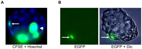
Figure 3. B10 cells contribute to the trophectodermal layer upon injection into rat blastocyst.
(A): Rat blastocyst containing CFSE- labelled cells in the trophectodermal layer (arrow), merged image of carboxyfluorescein succinimidyl ester (CFSE) fluorescence and Hoechst 33342 is shown, arrowhead indicates non-included cells in blastocoel. (B): Rat blastocyst with B10 cell expressing EGFP in the trophectoderm layer (arrow), fluorescent image and merged image of EGFP with differential interference contrast (Dic) is shown. Photos were taken with 40× objective.

