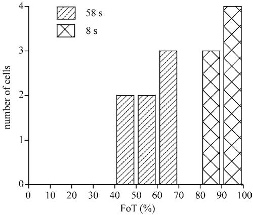
Figure 4.
Fraction of boundary with significant translocation (FoT). The FoT value is defined as the fraction of the boundary that shows a significantly higher fluorescence intensity than in the cytosol. The FoT values were determined for 7 cells at 8 and 58 s after stimulation. The FoT value at 8 s after stimulation is very high, indicating PHCrac-GFP associates to nearly the entire boundary, whereas at 58 s after stimulation PHCrac-GFP is found at only ∼50% of the boundary in patches.

