Figure 3.
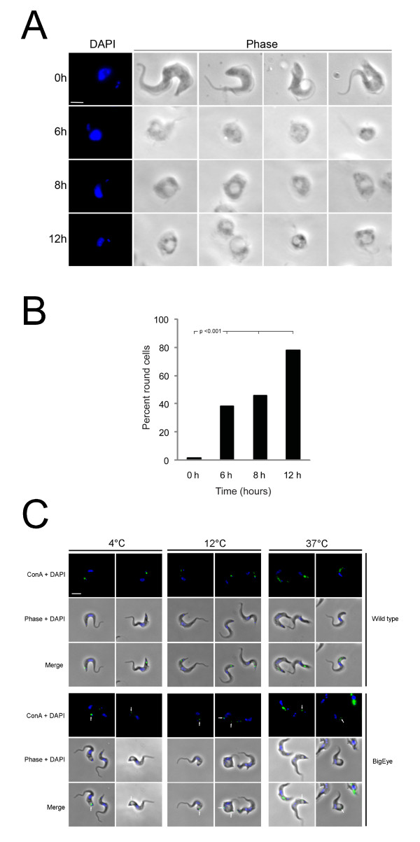
Reduced glucose levels in growth media for T. brucei leads to the appearance of the 'BigEye' phenotype and is lethal to BSF parasites. A. T. brucei BSF parasites were cultured in RPMI-1640 media containing 5 mM D-glucose and dialyzed (glucose-free) 10% FBS. At time zero the parasites have normal phenotype, but after six hours the cells became rounded. From six to twelve hours the flagellar pocket increased in diameter. Scale bar 2 um. B. The number of parasites with BigEye phenotype increases with time when grown in reduced glucose conditions. Cultures in glucose-replete medium contain essentially no BigEye cells, but by contrast, in reduced glucose medium (5 mM) at six hours rounded cells contribute 35% of the population, which increases to 45% and 80% by eight and twelve hours respectively. Statistical support for a significant increase in rounded cells is indicated using a chi-squared test. Note that at least 200 cells were examined for each condition, and all treated conditions are highly significantly different from the control (p < 0.001). C. Limiting glucose levels affects endocytosis in BSF parasites. ConA uptake was observed following growth under limiting glucose conditions. Cells in complete medium showed normal morphology while glucose-starved cells displayed the BigEye phenotype as observed by phase contrast. Location of FITC-ConA (green) in fixed cells co-stained with DAPI (blue) was observed by fluorescence microscopy at 4°C (flagellar pocket), 12°C (early endosomes) and 37°C (lysosomes). Arrows indicate ConA trapped at the flagellar pocket of BigEye cells. Scale bar is 2 um.

