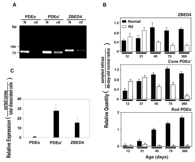
Figure 2.
Expression level of ZBED4 and cell-specific marker mRNAs in normal and rd mouse retinas and in cone-sorted cells. (A) RT-PCR amplification of 80-day-old normal and rd mouse retinal mRNAs using primer sets, PDEα (for PDEα) and PDEα′-1 (for PDEα′) and ZB27 (for ZBED4). The resulting RT-PCR products were separated on a 1.2% agarose gel and stained with ethidium bromide. ZBED4 is expressed in rd retina but at a lower level than in normal retina, similar to the expression of PDEα′, suggesting the presence of ZBED4 mRNA in cones but not ruling out its expression in the inner retina. (B) Quantitative real-time RT-PCR. Levels of ZBED4 mRNA in normal and rd mouse retinas at different times of postnatal development relative to those of 40-day-old normal retina were compared to the relative levels of cell-specific markers expressed in the same samples (normalized to β-actin mRNA level using primer set mA1; Table 1). Rod-specific marker: PDEα; cone-specific marker: PDEα′. (C) Relative expression of rod PDEα, cone PDEα′, and ZBED4 mRNA between flow cytometry–sorted mouse cones and dissociated retinal cells measured by QPCR using β-actin cDNA as normalizer. The primer sets are described in Table 1.

