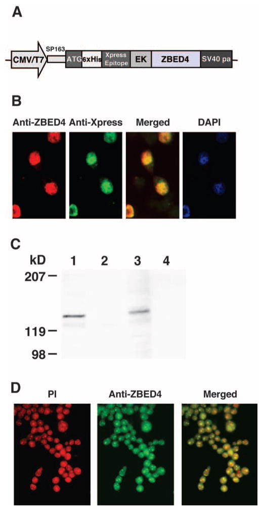
Figure 5.
Distribution of expressed ZBED4 in HEK293. (A) ZBED4 expression construct. (B) Immunocytochemical detection of the ZBED4 protein after transient transfection of the expression vector into HEK293 cells. Cells were double stained with antibodies against ZBED4 and Xpress and the resultant images were merged. Nuclear localization of expressed ZBED4 was detected by both antibodies and confirmed by staining with DAPI. (C) Western blot of nuclear and cytoplasmic extracts of HEK293 cells transfected for 24 or 72 hours. The blot was hybridized with anti-Xpress antibody. The expressed protein was present in the nuclear extract, confirming its immunocytochemical localization. Lanes 1 and 3: nuclear extracts; lanes 2 and 4: cytosolic extracts; lanes 1 and 2: extracts obtained after transfecting cells for 24 hours; lanes 3 and 4: extracts obtained after transfecting cells for 72 hours. (D) Localization of ZBED4 protein in Y79 cells. Note the granular pattern of ZBED4 staining in the nuclei.

