Figure 6.
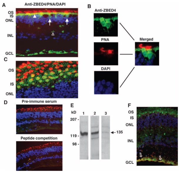
Localization of ZBED4 in human retina. (A, C) Human retinal sections were double-stained with N terminus ZBED4 antibody followed by FITC-conjugated secondary antibody (green), rhodamine-conjugated PNA (red), and DAPI (blue). (A) Cone nuclei (arrows) and inner segments (arrowheads) are stained green, and the cone matrix is stained red. Note also the anti-ZBED4 staining of the innermost layer of the retina and of the cone pedicles (open arrowhead). (B) Magnified images individually stained with anti-ZBED4, PNA, and DAPI with the use of appropriate filters, and the merging of the three images. (C) An obliquely cut retinal section shows the cone inner segment localization of ZBED4 surrounded by PNA-stained cone extracellular matrix. OS, outer segments; IS, inner segments; ONL, outer nuclear layer; INL, inner nuclear layer; GCL, ganglion cell layer. (D) Top: section incubated with rabbit preimmune serum, rhodamine-conjugated PNA and DAPI. Bottom: section incubated with ZBED4 antibody that had been absorbed with the ZBED4 peptide used to generate it, rhodamine-conjugated PNA and DAPI. The absence of ZBED4 staining validates the ZBED4 antibody specificity. (E) Western blot of proteins from the nuclear extract of mouse thymus (lane 1) and from the nuclear (lane 2) and cytosolic (lane 3) fractions of human retina. ZBED4 (arrow) is very abundant in mouse thymus nuclei; therefore, it was used as a positive control in all Western blot analyses. (F) An obliquely cut human retinal section double-labeled with rabbit polyclonal anti-ZBED4 (green) and anti-human vimentin (red); nuclei were stained with DAPI (blue). Arrows: colocalization of vimentin and ZBED4 in Müller cell endfeet. Magnification: (A, D, F) ×400; (C) ×600.

