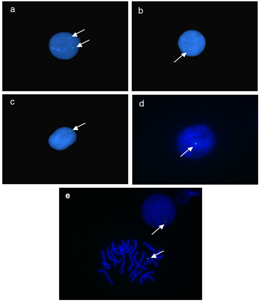
Fig. 4.
Fluorescence in situ hybidization (FISH) of cells harvested from pT2βGFP transgenic Xenopus tropicalis. Interphase nuclei were prepared from circulating blood cells harvested from individual tadpoles and probed with FITC-labled GFP for detection. White arrows indicate location of the GFP probe in the samples. a: pT2βGFP X. tropicalis founder line 4M. b: pT2βGFP founder line 5M c: pT2βGFP founder line 6M. d: pT2βGFP founder line 7M. e: Interphase nuclei and metaphase spread of founder line 7M.

