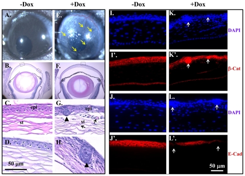
Fig. 5.
Expression of ΔE3β-catenin induced corneal epithelial hyperplastic nodules and downregulated E-cadherin. (A-H) Stereo micrographs of ocular surface in Krt12rtTA/Wt/tetO-Cre/Ctnnb1floxE3/Wt mouse at P21 induced without (A) or with (E) Dox since E14.5. It was obvious that ΔE3β-catenin expression led to corneal opacity and profound neovascularization (arrows in E). H&E staining showed that non-induced eye developed into a well-organized stratified corneal epithelium (B,C,D). However, in Dox-treated eye, corneal epithelial cells arranged into epithelial nodules instead of stratified epithelium (arrowhead in G,H), thereby corneal integrity was compromised concomitant with angiogenesis (arrows in G). (I-L′) Corneal sections from P21 of Krt12rtTA/Wt/tetO-Cre/Ctnnb1floxE3/Wt mice induced without Dox (I,I′,J,J′) or with (K,K′,L,L′) were subjected to immunofluorescence staining of β-catenin (I,I′,K,K′) and E-cadherin (J,J′,L,L′). Note that ΔE3β-catenin was accumulated in the nucleus (arrows in K′) and E-cadherin expression was reduced in the epithelial nodules (arrows in L′). ep, epithelium; st, stroma.

