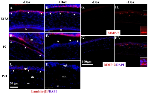
Fig. 6.
Expression of ΔE3β-catenin disrupted basement membrane formation concomitantly with upregulated MMP-7. (A-F) Corneal sections from E17.5 (A,D), P2 (B,E), and P21(C,F) of Krt12rtTA/Wt/tetO-Cre/Ctnnb1floxE3/Wt triple-transgenic mice induced without Dox (−Dox) or with (+Dox) were subjected to immunofluorescence staining of laminin-β1. In non-induced mice, laminin-β1 expression was detected in basolateral region at E17.5 (A) and P2 (B) and more restricted to the basal region at P21(C), respectively, of the corneal epithelial cells. Note that, in Dox-treated cornea, the laminin-β1 expression pattern did not seem to be changed at E17.5 (D) but was drastically downregulated in P2 (E) and undetectable in P21 (F) (arrows in D,D′,E,E′). (G-H′) Corneal sections from P21 of Krt12rtTA/Wt/tetO-Cre/Ctnnb1floxE3/Wt mice induced without Dox (G,G′) or with (H,H′) were subjected to immunofluorescence staining of MMP-7. Note that MMP-7 expression level was undetectable in the normal corneal epithelium (G,G′) but upregulated by ΔE3β-catenin in epithelial cells (H,H′). epi, epithelium; str, stroma; en, endothelium.

