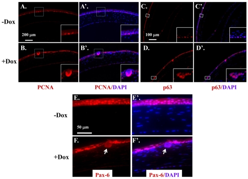
Fig. 7.
Expression of ΔE3β-catenin in corneal epithelium increased cell proliferation, upregulated p63 expression but downregulated Pax-6. (A-F′) Corneal sections from P21 of Krt12rtTA/Wt/tetO-Cre/Ctnnb1floxE3/Wt mice induced without Dox (−Dox) or with (+Dox) were subjected to immunofluorescence staining of PCNA (A,A′,B,B′), p63 (C,C′,D,D′), and Pax-6 (E,E′,F,F′), respectively. PCNA was highly expressed in the epithelial nodules of Dox-treated mice (insets of B,B′). Likewise, p63 expression was restricted to the epithelial basal cells (insets of C,C′) in non-induced mice but was detected in the entire epithelial nodules of Dox-treated mice (insets of D,D′). Pax-6 was expressed in the full thickness of epithelium of non-induced mice (E,E′). Interestingly, Pax-6 expression in Dox-treated cornea was comparable with that in non-induced mice and diminished only in the epithelial nodules (arrows in F,F′).

