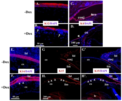
Fig. 8.
Expression of ΔE3β-catenin altered keratin expression pattern in corneal epithelium. (A-H′) Corneal sections from P21 of Krt12rtTA/Wt/tetO-Cre/Ctnnb1floxE3/Wt mice induced without Dox (−Dox) or with (+Dox) were subjected to immunofluorescence staining of K12 (A,B) K4 (C,D), K14 (E,F), and K15 (G,G′,H,H′). K12 was expressed in the full-thickness of epithelium (A) of non-induced mice. However, K12 expression was dramatically reduced in Dox-induced epithelial nodules (arrows in B). However, K4 expression pattern was not altered by ΔE3β-catenin (C,D), but K15 was expressed in limbal and conjunctival epithelia but not cornea of the non-induced mice (E,E′). Interestingly, ΔE3β-catenin expression showed extension of K15 expression to the central cornea (arrows in F,F′). Epithelial nodules were stained positive for K14 (arrows in H) and might assume epithelial progenitor cell phenotype.

